Database content
Identifying the entirety of Human leukocyte antigen (HLA) presented peptides are indispensable for the development of cancer immunotherapies, defined as the immunopeptidome. Epitopes derived from noncoding regions expand immunopeptidome in cancer. Here, we developed the IEAtlas database to provide an atlas of HLA-presented immunogenic epitopes derived from non-coding regions.
IEAtlas:
I) re-analyzes public HLA immunopeptidome mass spectrometry data from 2,747 samples of 15 cancer types and 30 non-cancerous tissues
II) constructs a theory library including 560112 translated ORFs from non-coding regions
III) detects a total of 145366 and 100504 non-canonical epitopes binding by HLA I and II respectively.
IV) queries the predicted information of both presentation features and recognition features
V) provides the clinical relevance of corresponding genes
Recent evidence highlights the importance of non-canonical epitopes, which can also elicit anti-tumor immune responses and mainly originated in translation products of transcripts from putative non-protein-coding regions, including the 5′ and 3′ untranslated regions (UTRs), overlapping yet out-of-frame alternative ORFs in annotated protein-coding genes, long non-coding RNAs (lncRNAs), pseudogenes and so on. ORFs derived from putative non-protein-coding regions were defined as non-canonical ORFs (ncORFs). We integrated Ribo-seq supported ncORFs from RPFdb (1), nuORFdb (2) and Translnc (3) with their basic annotations.
All the available MS-based immunopeptidome datasets were obtained from commonly used proteome databases, including PRIDE (4), MassIVE.quant (5), PeptideAtlas (6), JPOST (7), Panorama (8) and iProX (9). Currently, IEAtlas reanalyzed 2,747 samples of 15 cancer types and 30 non-cancerous tissues against the integrated benchmarked theory library of non-canonical peptides.
Information of MS-based immunopeptidome datasets
| Tissue |
Type |
HLA Type |
Resource |
IEAtlas further evaluates the immunogenicity via several commonly used immunogenic features, including HLA-binding affinity, stability, and TCR recognition(10-13). Epitopes with MHC binding affinity < 500 nM, MHC binding stability > 1.4 h, and T cell recognition probability > were defined as immunogenic epitopes.
To provide a convenient way for users to investigate the functions of epitopes and genes of interest, IEAtlas has also developed five flexible tools.
Including:
1. Epitopes-in-Cancer provides epitopes detected by MS-based immunopeptidome across different cancer.
2. Epitopes-in-Normal-Tissues provides epitopes detected by MS-based immunopeptidome across different normal tissues.
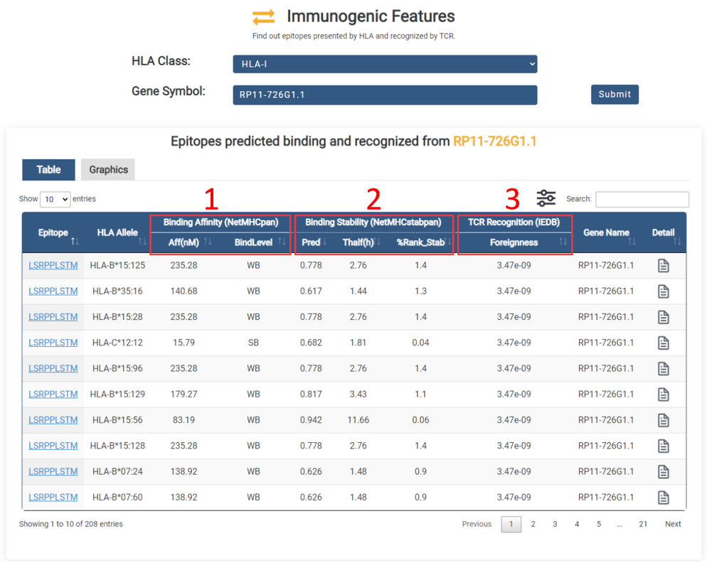
3. Immunogenic-Features provides the predicted information of both presentation features and recognition features of immunogenic epitopes.
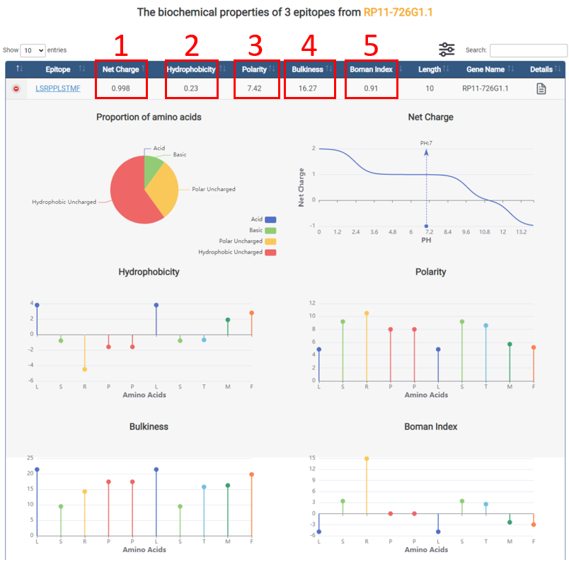
4. Biochemical-Properties provides the biochemical properties of all epitopes from cancer and normal tissues.
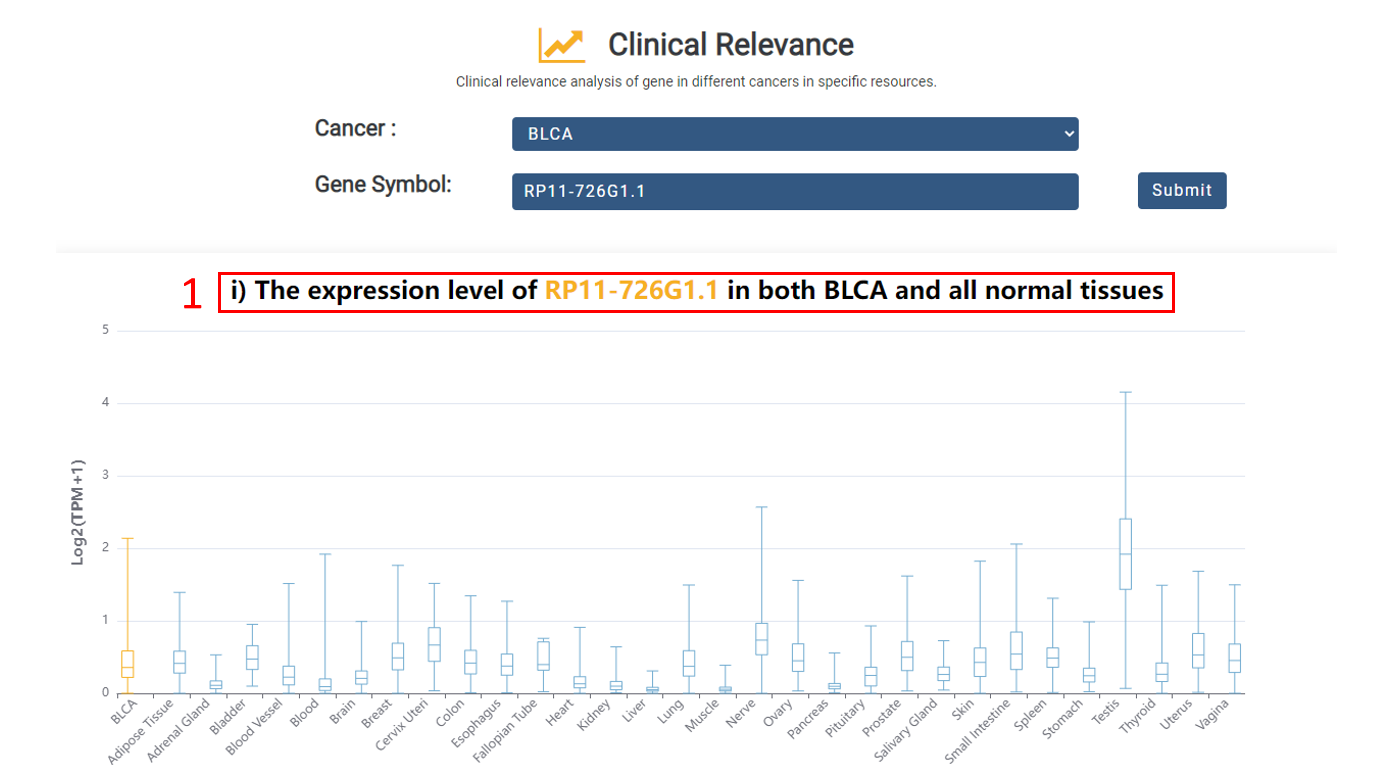
5. Clinical-Relevance performs differential expression analysis and survival analysis for corresponding genes.
Web interface
IEAtlas provides a user-friendly searching and browsing interface.
1. Main functions of the database are provided in menu bar form.
2. Click "Quick Search" button to start a quick search.
1. Select normal tissues or cancers.
2. Select tissue type.
3. Input gene symbol.
4. Select HLA class(HLA-I or HLA-II).
5. Input HLA allele.
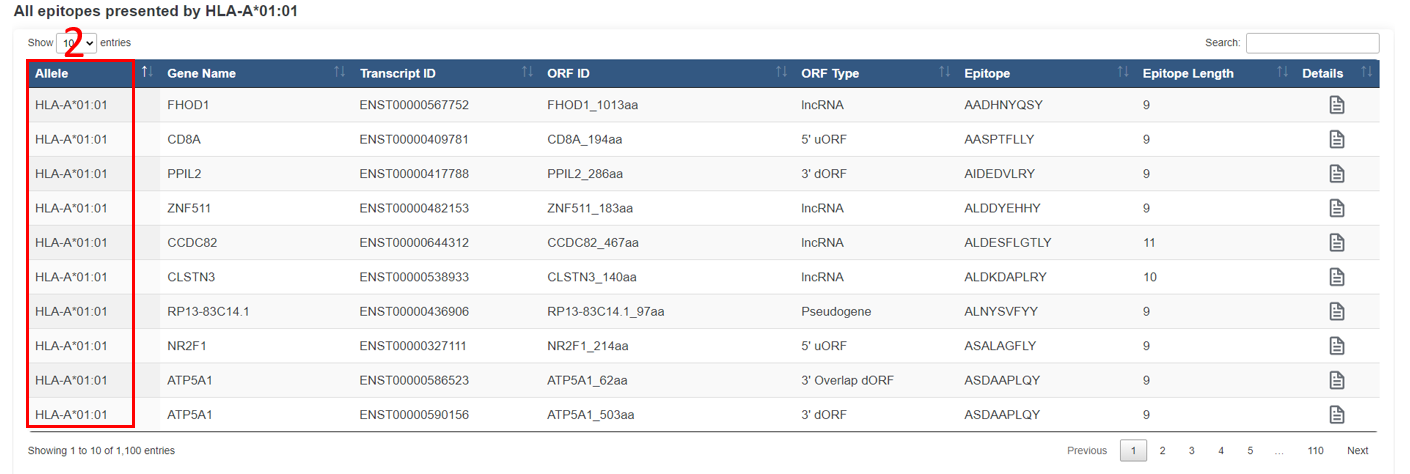
For the browse page, we provide all epitopes indentified by MS-based immunopeptidome across all tissues in IEAtlas.
1. The size of the dot indicates the number of epitopes in the tissue.
2. The color of the dot indicates tissue type.
3. Click to browse all epitopes in specific tissue.
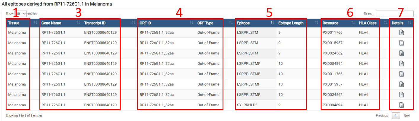
The result page of epitopes is displayed as below.
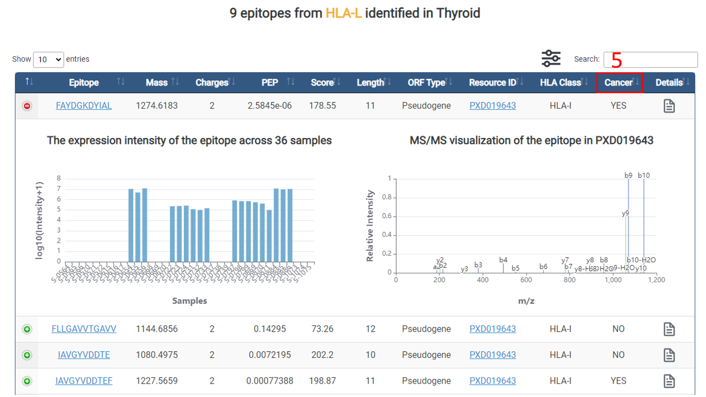
1. Tissue.
2. HLA allele.
3. Gene basic information.
4. ORF basic informaton.
5. Epitope basic informaton.
6. Resource.
7. Click to view the detail information.
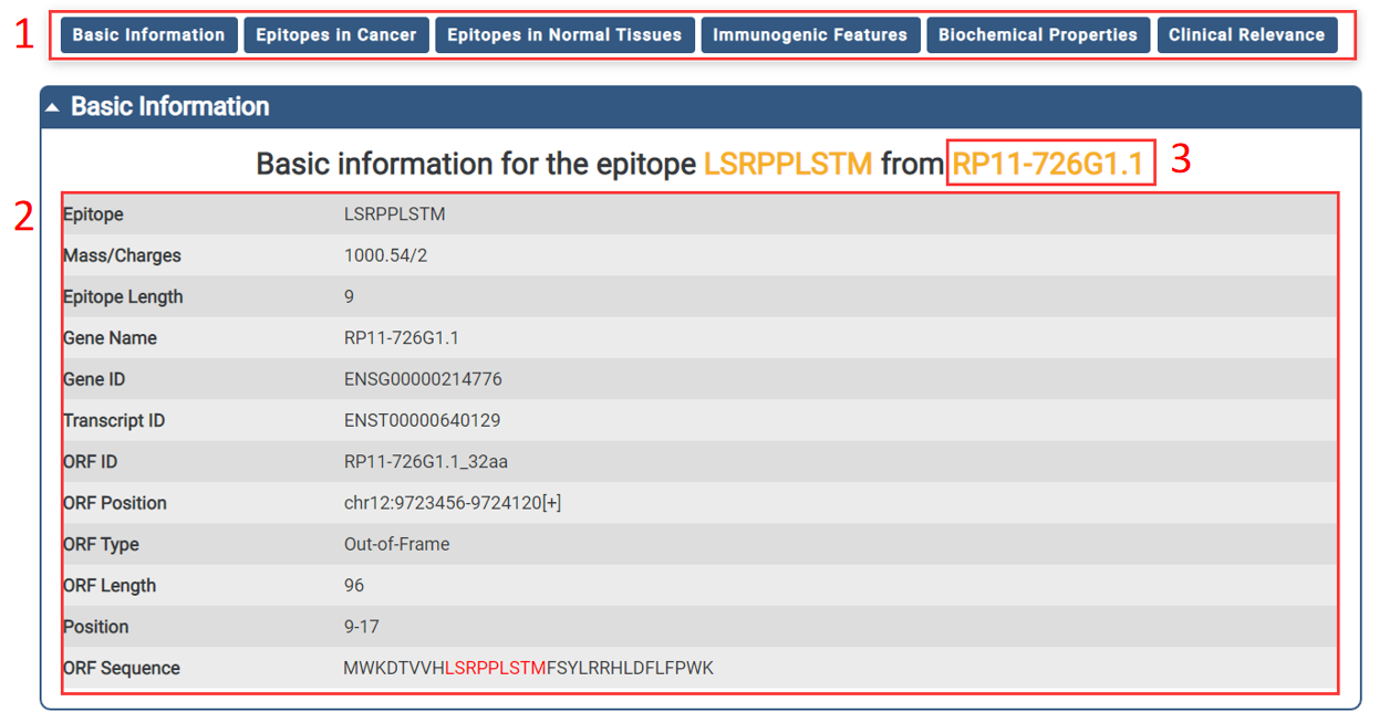
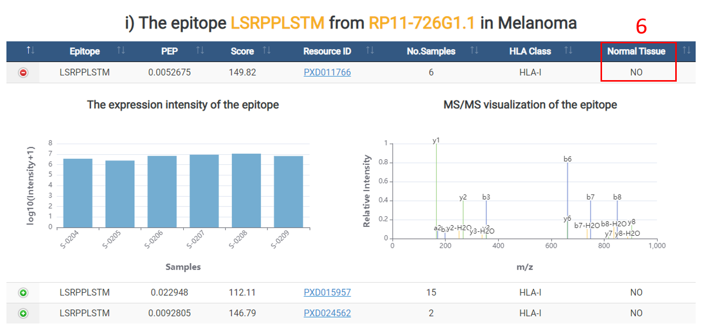
For each epitope, we provide basic information, which is displayed as below.
1. You could directly go to the module of interest by click the axis.
2. The basic information of the epitope.
3. Click the gene symbol for linking the corresponding Ensembl page.
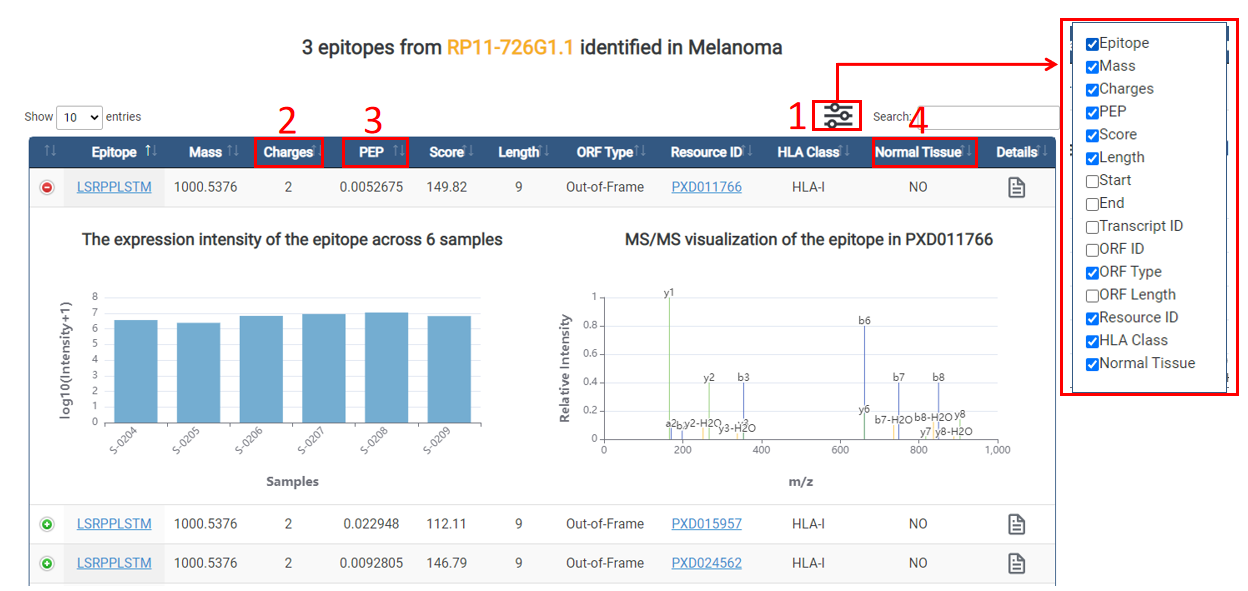
1. Browse the hidden columns.
2. PEP: posterior error probability of the identification. This value essentially operates as a p-value, where smaller is more significant.
3. Score: andromeda score for the best associated MS/MS spectrum.
4. No.Samples: the number of samples in the resource.
5. Cancer: whether the epitopes occur in cancers or not.
6. No.Samples: the number of samples in the resource.
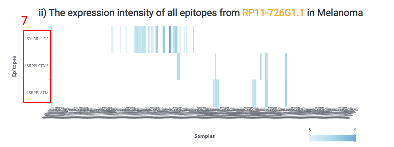
7. All epitopes derived from the same gene in cancer of interest.
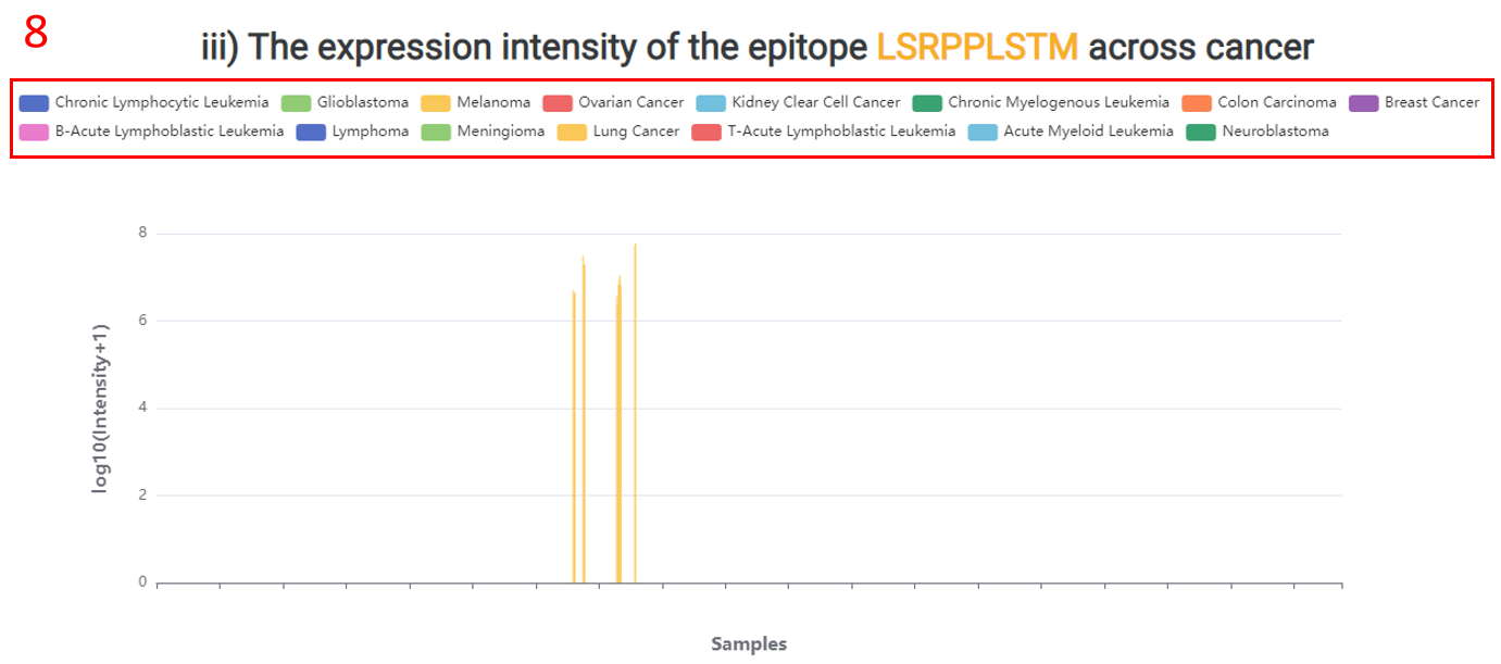
8. The expression intensity of the epitope across all cancers.
Both presentation features and recognition features of all immunogenic epitopes.
1. Epitope binding affinity was predicted by NetMHCpan and NetMHCIIpan with default settings(either as a strong-binding threshold of 50nm or weak-binding threshold of 500nm).
2. Epitope binding stability was predicted by NetMHCstabpan using default parameters(binding stability greater than 1.4 h).
3. Foreignness: TCR recognition probability derived from homology to known pathogenic peptides in IEDB using the multistate thermodynamic model described by Luksza et al(foreignness more than 1e-16).
4. Click to enter the AFND database.
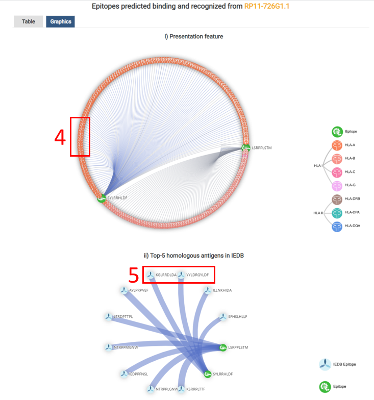
5. Click to the IEDB database. According to the sequence alignment scores, the top-5 homologous antigens in IEDB were also shown as a network.
In this page, we provide biochemical properties of all epitopes.
1. Net Charge of epitopes when pH=7.
2. Mean hydrophobicity or epitopes.
3. Mean polarity or epitopes.
4. Mean bulkness or epitopes.
5. Boman Index computes the potential protein interaction index proposed by Boman (2003) based in the amino acid sequence of a protein.
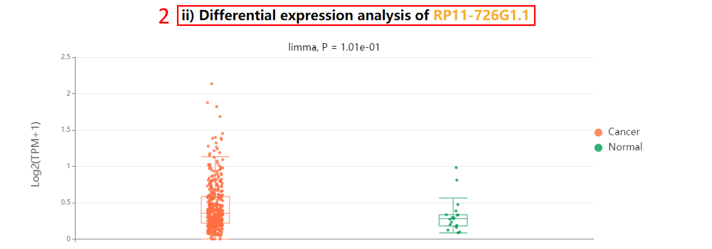
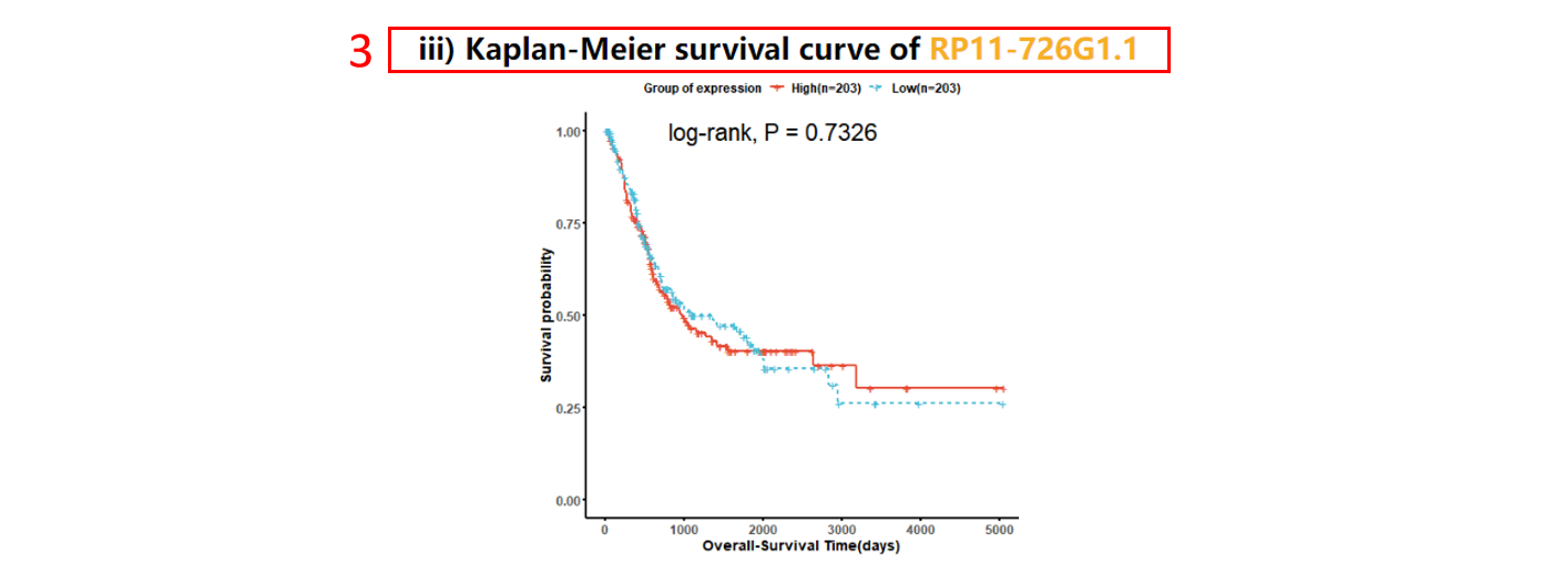
For each gene, we provide differential expression analysis and survival analysis across 33 cancer types in TCGA and across 30 normal tissues in GTEx.
1. Gene expression in TCGA tumor samples vs GTEx.
2. Differential expression analysis in TCGA.
3. Survival analysis.
1. Wang, H., Yang, L., Wang, Y., Chen, L., Li, H. and Xie, Z. (2019) RPFdb v2.0: an updated database for genome-wide information of translated mRNA generated from ribosome profiling. Nucleic Acids Res, 47, D230-D234.
2. Ouspenskaia, T., Law, T., Clauser, K.R., Klaeger, S., Sarkizova, S., Aguet, F., Li, B., Christian, E., Knisbacher, B.A., Le, P.M. et al. (2022) Unannotated proteins expand the MHC-I-restricted immunopeptidome in cancer. Nat Biotechnol, 40, 209-217.
3. Lv, D., Chang, Z., Cai, Y., Li, J., Wang, L., Jiang, Q., Xu, K., Ding, N., Li, X., Xu, J. et al. (2022) TransLnc: a comprehensive resource for translatable lncRNAs extends immunopeptidome. Nucleic Acids Res, 50, D413-D420.
4. Perez-Riverol, Y., Bai, J., Bandla, C., Garcia-Seisdedos, D., Hewapathirana, S., Kamatchinathan, S., Kundu, D.J., Prakash, A., Frericks-Zipper, A., Eisenacher, M. et al. (2022) The PRIDE database resources in 2022: a hub for mass spectrometry-based proteomics evidences. Nucleic Acids Res, 50, D543-D552.
5. Choi, M., Carver, J., Chiva, C., Tzouros, M., Huang, T., Tsai, T.H., Pullman, B., Bernhardt, O.M., Huttenhain, R., Teo, G.C. et al. (2020) MassIVE.quant: a community resource of quantitative mass spectrometry-based proteomics datasets. Nat Methods, 17, 981-984.
6. Schwenk, J.M., Omenn, G.S., Sun, Z., Campbell, D.S., Baker, M.S., Overall, C.M., Aebersold, R., Moritz, R.L. and Deutsch, E.W. (2017) The Human Plasma Proteome Draft of 2017: Building on the Human Plasma PeptideAtlas from Mass Spectrometry and Complementary Assays. J Proteome Res, 16, 4299-4310.
8. Sharma, V., Eckels, J., Taylor, G.K., Shulman, N.J., Stergachis, A.B., Joyner, S.A., Yan, P., Whiteaker, J.R., Halusa, G.N., Schilling, B. et al. (2014) Panorama: a targeted proteomics knowledge base. J Proteome Res, 13, 4205-4210.
9. Ma, J., Chen, T., Wu, S., Yang, C., Bai, M., Shu, K., Li, K., Zhang, G., Jin, Z., He, F. et al. (2019) iProX: an integrated proteome resource. Nucleic Acids Res, 47, D1211-D1217.
10. Wells, D.K., van Buuren, M.M., Dang, K.K., Hubbard-Lucey, V.M., Sheehan, K.C.F., Campbell, K.M., Lamb, A., Ward, J.P., Sidney, J., Blazquez, A.B. et al. (2020) Key Parameters of Tumor Epitope Immunogenicity Revealed Through a Consortium Approach Improve Neoantigen Prediction. Cell, 183, 818-834 e813.
11. Luo, X., Huang, Y., Li, H., Luo, Y., Zuo, Z., Ren, J. and Xie, Y. (2022) SPENCER: a comprehensive database for small peptides encoded by noncoding RNAs in cancer patients. Nucleic Acids Res, 50, D1373-D1381.
12. Chen, J., Brunner, A.D., Cogan, J.Z., Nunez, J.K., Fields, A.P., Adamson, B., Itzhak, D.N., Li, J.Y., Mann, M., Leonetti, M.D. et al. (2020) Pervasive functional translation of noncanonical human open reading frames. Science, 367, 1140-1146.
13. Bulik-Sullivan, B., Busby, J., Palmer, C.D., Davis, M.J., Murphy, T., Clark, A., Busby, M., Duke, F., Yang, A., Young, L. et al. (2018) Deep learning using tumor HLA peptide mass spectrometry datasets improves neoantigen identification. Nat Biotechnol.